The physical therapist can use ultrasound as a supplementary examination tool in addition to the regular physical examination and intake interview, in order to make a diagnosis or to rule out certain aspects. The ultrasound examination enables the physical therapist to make a better diagnosis and thus draw up a more specialized treatment plan.
What symptoms warrant an ultrasound?
Do you suffer from one or more of the following symptoms? If so, the physical therapist may decide to use ultrasound. This is up to the physical therapist to determine.
• Shoulder: bursitis, tendon and muscle damage/tears, bone abnormalities, and calcifications
• Elbow: tennis elbow, golfer's elbow, and tendon damage
• Wrist: carpal tunnel syndrome, tendon damage, and small fissures or avulsion fractures
• Hip: bursitis, capsule and tendon damage
• Knee: hamstring, knee ligaments and tendon damage
• ankle: joint, ankle ligaments, tendon damage and small fissures or avulsion fractures
• Achilles tendon: tears and inflammation
• General: muscle tears and tendon damage
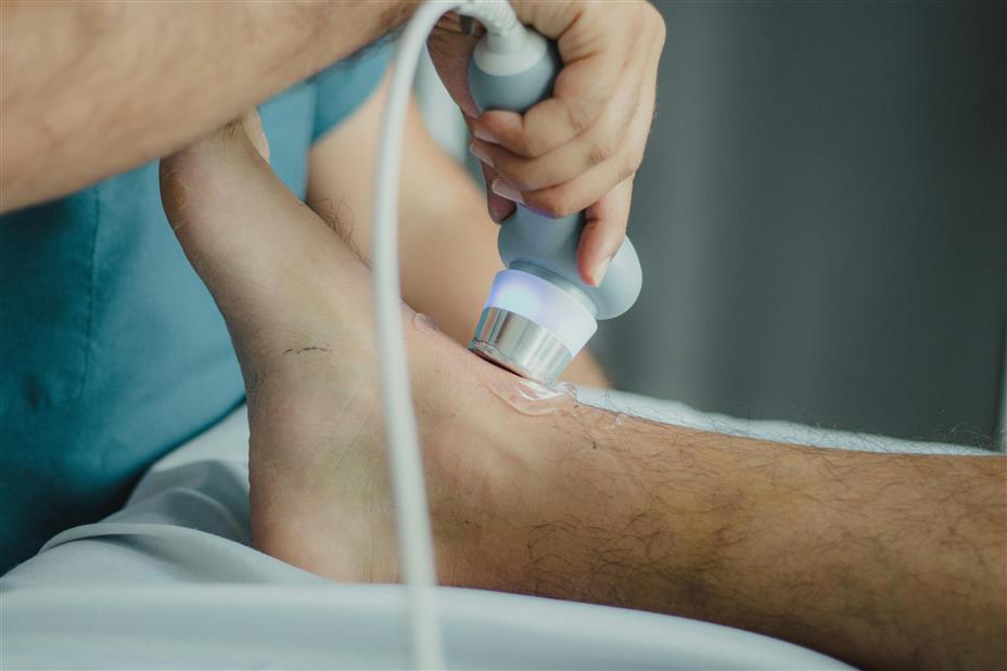
How does ultrasound work?
Ultrasound uses high-frequency sound waves that are inaudible to the human ear. The sound waves are sent into the body by placing a transducer (small scanner, probe) directly on the skin. The transducer is a device that can send and receive sound waves. To ensure that the sound is received and transmitted properly, gel is applied between the transducer and the skin. The transmitted sound is reflected back by the organs/bones in the body. The reflected sound is converted into an electrical signal by a computer, creating an image on the monitor. This image can be assessed by the physical therapist.
Making a diagnosis
Ultrasound is intended to make a diagnosis, but it also gives us insight into the direction of the treatment plan and the prognosis. It can also be used to effectively decide when other care providers should be called in if we, as physical therapists, are not the appropriate care providers to treat a complaint.